
Lunedì 17 Novembre 2025 Inizio Ore: 11:00 - Giornata intera
CONVEGNO
RI-VEDERE IL FUTURO: Genetica, Visione Artificiale e Nuove Tecnologie per Combattere la Cecità
MACULA TODAY 2025
Rome Cavalieri Waldorf Astoria - Sala Terrazza "Monte Mario"
Via Alberto Cadlolo 101, Roma
A causa dell’elevato numero di richieste, non è più possibile effettuare la prenotazione per la partecipazione in presenza.
Sarà comunque possibile seguire il convegno da remoto tramite collegamento Zoom.
PARTECIPA VIRTUALMENTE AL CONVEGNO
Convegno Macula Today 2025
RI-VEDERE IL FUTURO:
Genetica, Visione Artificiale e Nuove Tecnologie per Combattere la Cecità
Macula Today è il Convegno annuale organizzato dalla Macula & Genoma Foundation, Fondazione no profit che promuove la ricerca nell’ambito delle malattie oculari.
La Macula & Genoma Foundation si prefigge di promuovere una cultura della solidarietà sociale mediante la divulgazione e l’applicazione delle più recenti scoperte scientifiche, senza fini di lucro e senza barriere geografiche, sociali ed economiche.
L’edizione di quest’anno ospiterà un incontro del gruppo di interesse speciale (Special Interest Group Meeting) dedicato al tema del rimodellamento retinico interno, un’area di ricerca sempre più cruciale per la selezione dei pazienti e la definizione dei tempi ottimali per l’introduzione di nuove terapie, tra cui la terapia genica, la terapia cellulare, l’optogenetica, l’optopharmacologia e le protesi retiniche e corticali.
Le finalità del Macula Today
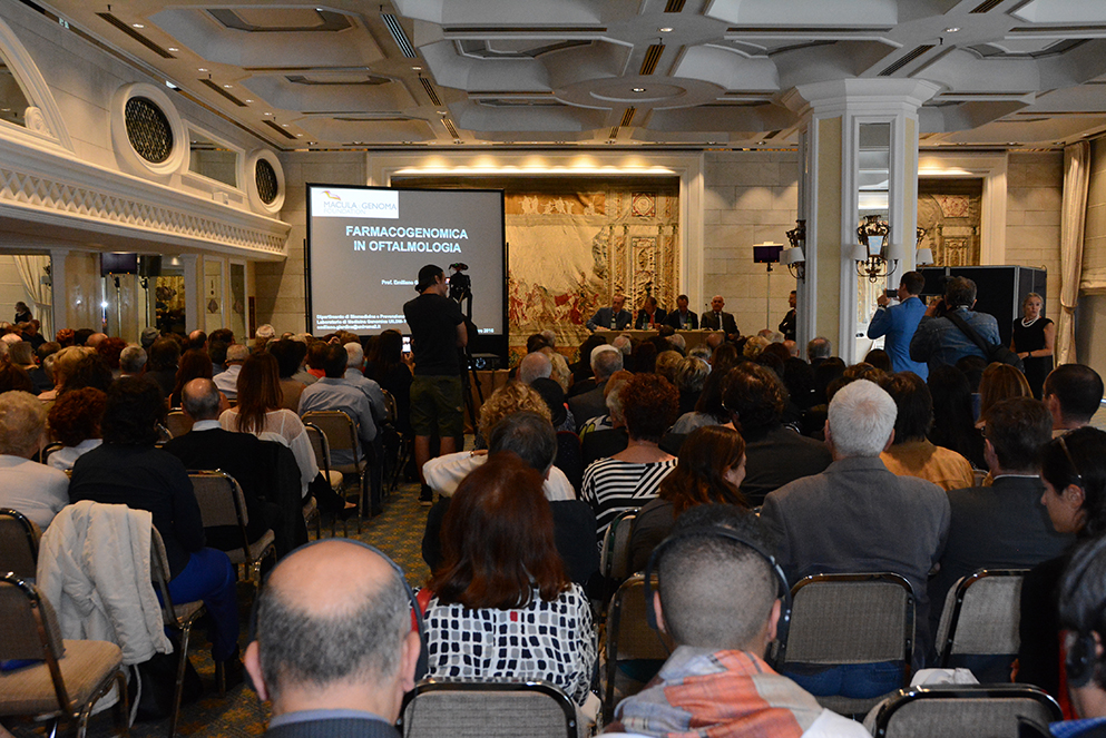
Il Convegno ospita ogni anno eminenti esperti del settore dell’oftalmologia per presentare i dati delle loro ricerche, tra le più innovative a livello mondiale. L’evento è rivolto non solo agli oculisti ma anche ai pazienti e ai non addetti ai lavori, permettendo a tutti, mediante l’utilizzo di un linguaggio semplice e chiaro, ma scientificamente corretto, di entrare in diretto contatto con il mondo della ricerca più all’avanguardia. Il Macula Today permette di essere informati su nuove terapie e traguardi terapeutici già disponibili o che potrebbero diventare tali nel prossimo futuro.
I relatori di questa edizione
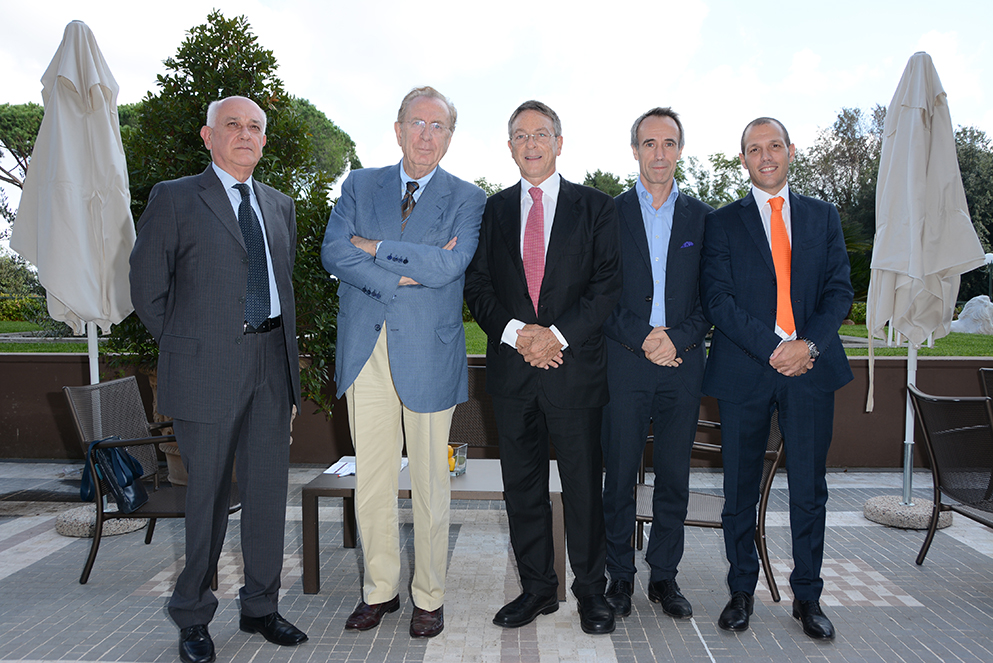
Quest’anno “Macula Today” ha il privilegio di ospitare nove ricercatori provenienti da eminenti università e istituti di ricerca:
-
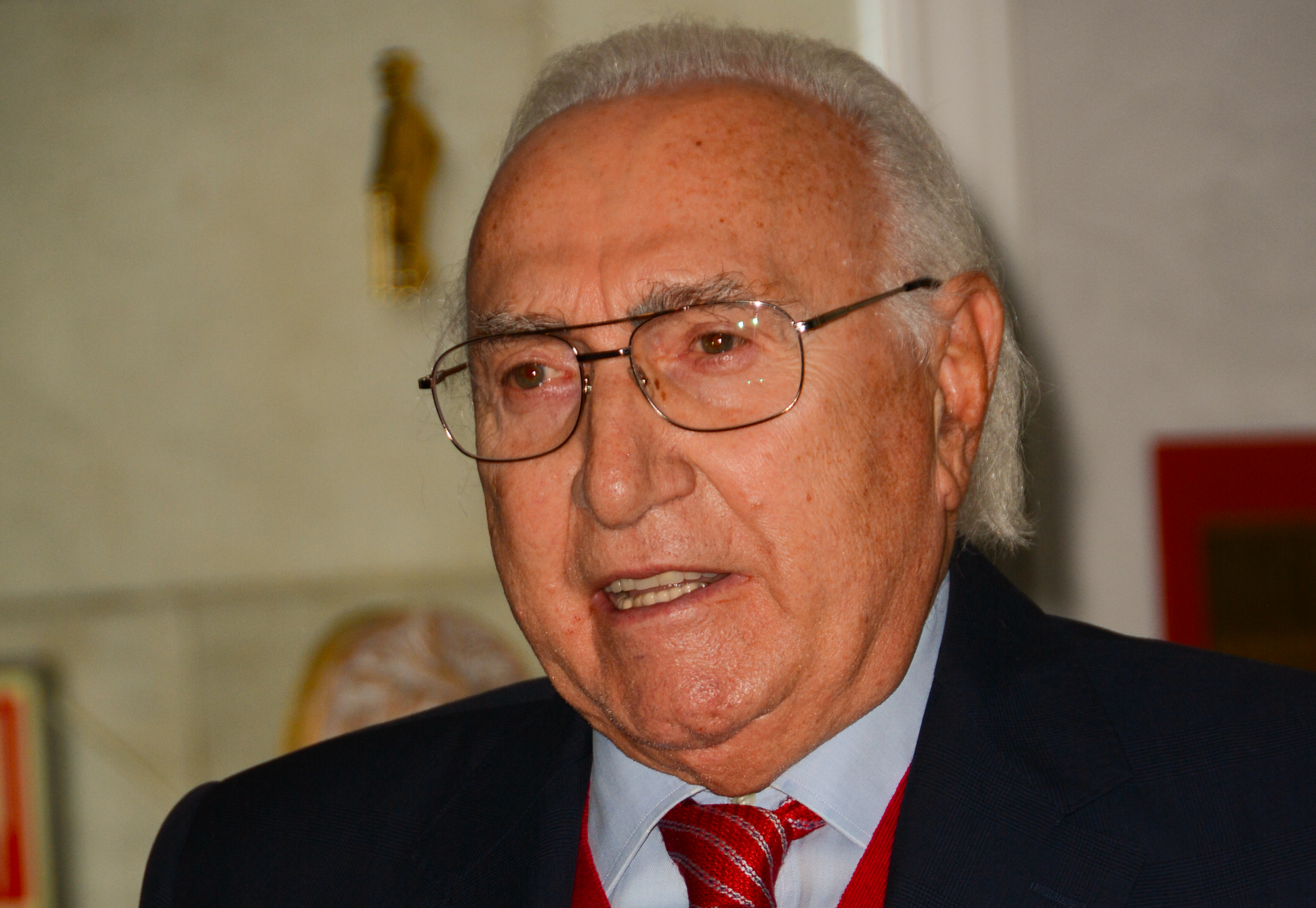
- Prof. Andrea Cusumano
Università degli Studi di Roma “Tor Vergata”, Roma; Rheinische Friedrich-Wilhelms-Universität, Bonn; Weill Cornell Medical College, New York
President, Macula & Genoma Foundation
- Prof. Andrea Cusumano
-
- Prof. Benedetto Falsini
Macula & Genoma Foundation, New York; Istituto di Oftalmologia, Università Cattolica del Sacro Cuore, Roma; Fondazione Policlinico Universitario A. Gemelli, Roma
- Prof. Benedetto Falsini
-
- Prof. Eduardo Fernández
Director, Instituto de Bioingeniería, Edificio Vinalopó, Universidad Miguel Hernández, Elche (Alicante), Spagna
- Prof. Eduardo Fernández
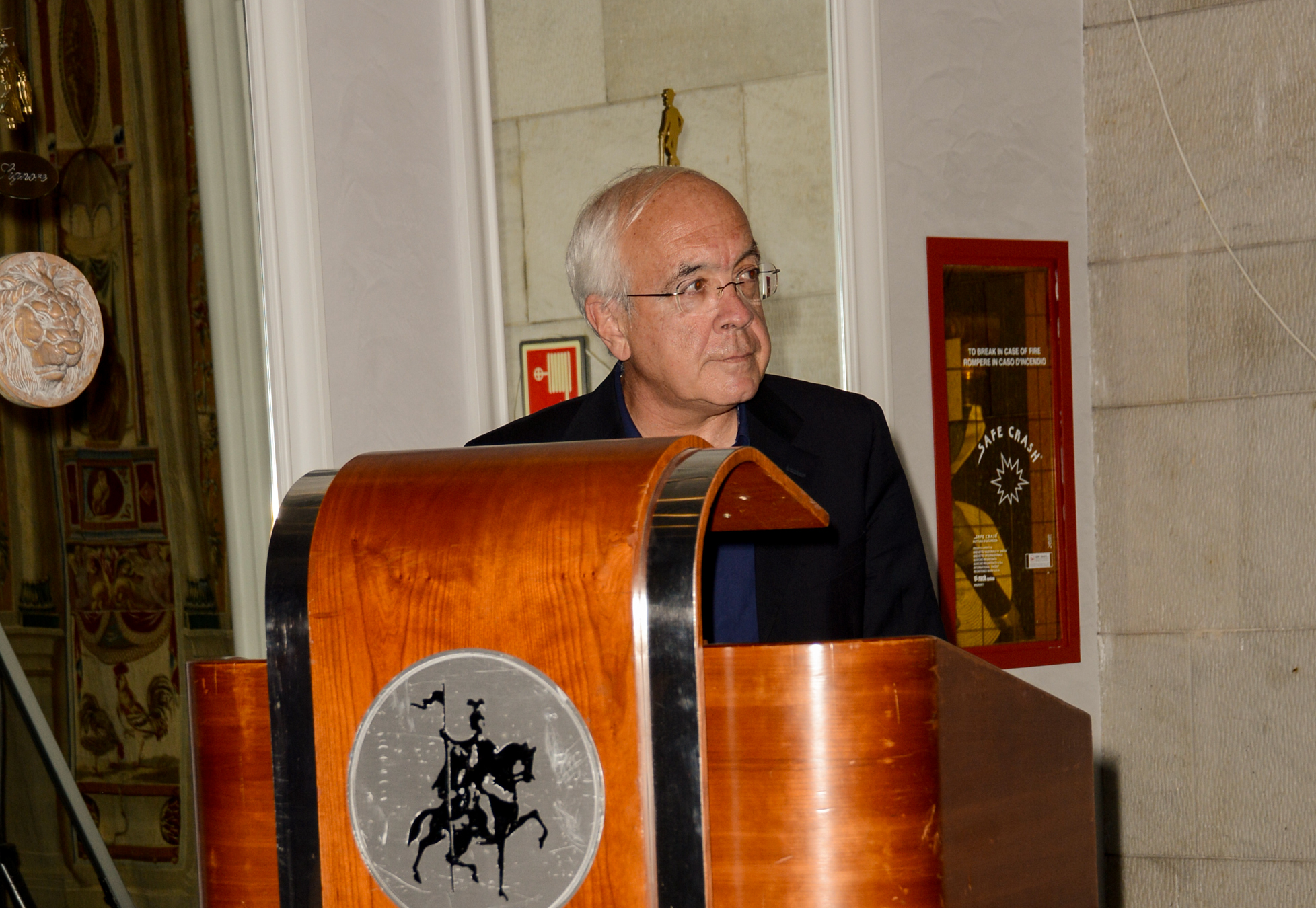
- Prof. Emiliano Giardina
Università degli Studi di Roma “Tor Vergata”, Roma; Director, Laboratorio di Medicina Genomica UILDM, Fondazione Santa Lucia, Roma
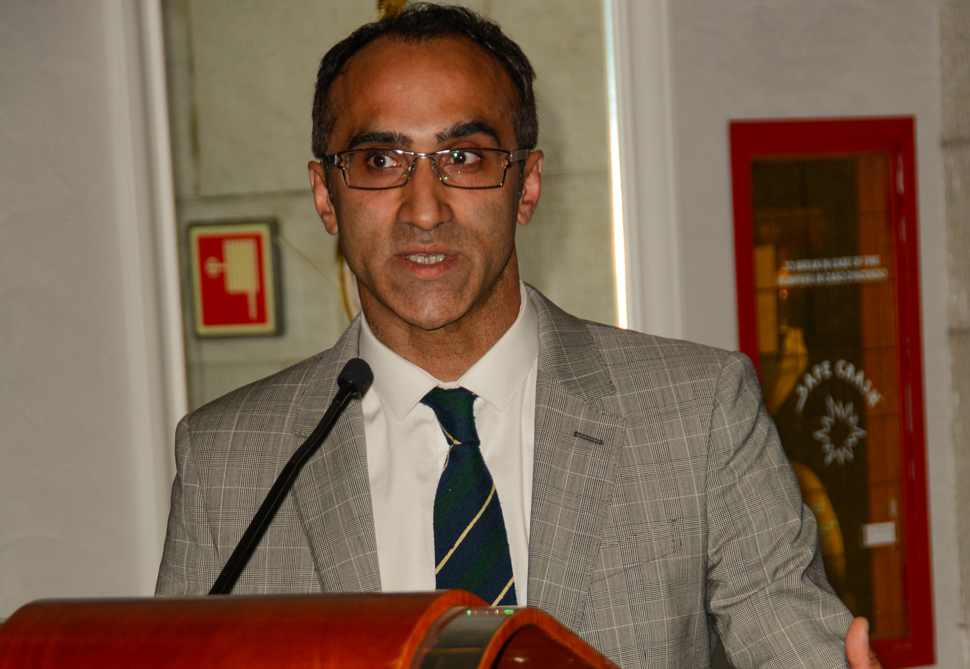
- Prof. Aniz Girach
Chief Medical Officer, SpliceBio, Barcellona, Spagna; Visiting/Honorary Professor, Wills Eye Hospital, Philadelphia, USA
-
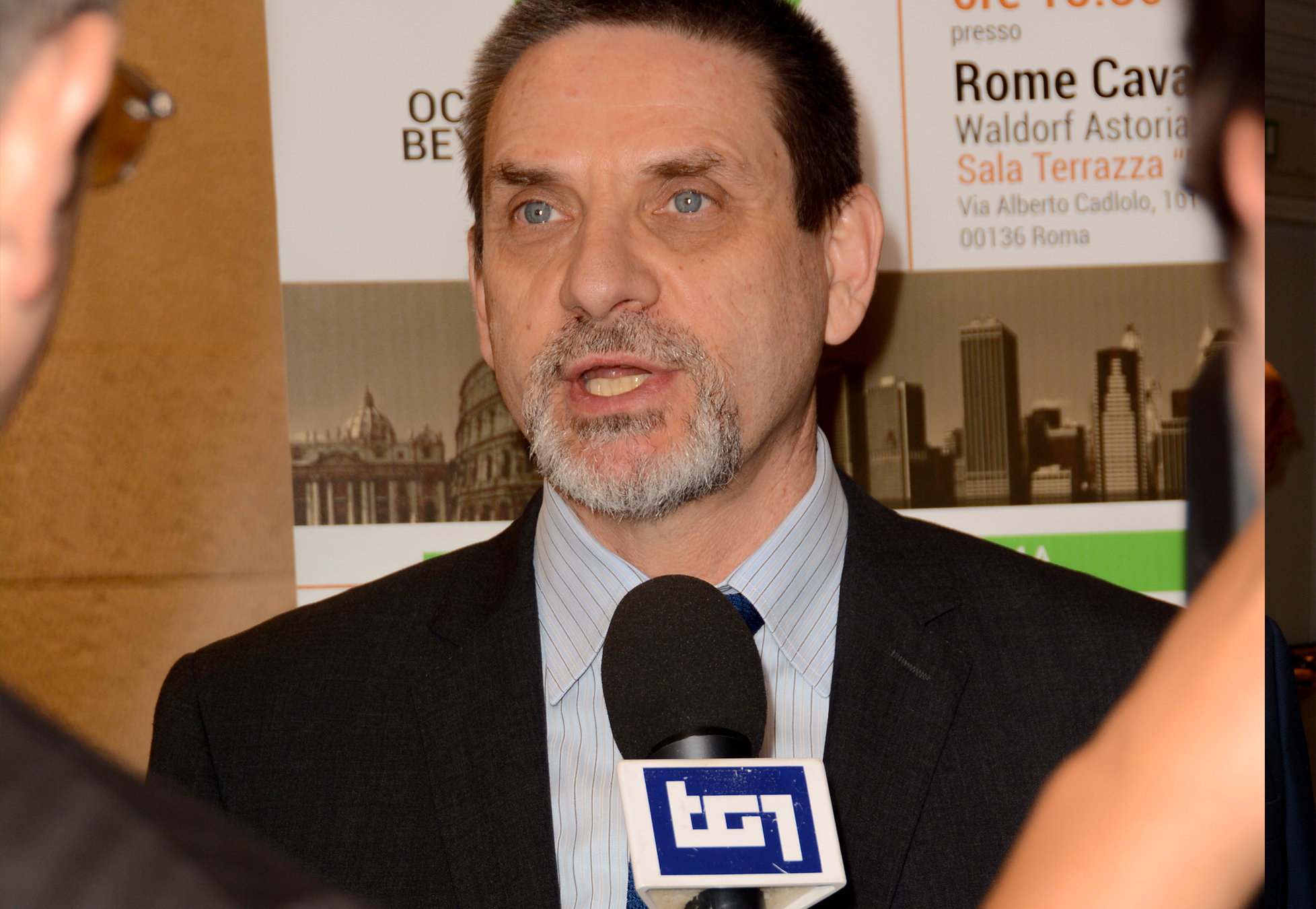
- Prof. Michael B. Gorin
Professor, Department of Ophthalmology, Retina and Vision Science Divisions; Harold and Pauline Price Chair in Ophthalmology, UCLA Jules Stein Eye Institute, California, USA
- Prof. Michael B. Gorin
-
- Prof. Richard H. Kramer
CH and Annie Li Chair in Molecular Biology of Diseases; Professor of Neurobiology, University of California, Berkeley, USA
- Prof. Richard H. Kramer
-
- Prof. Daniel Palanker
Professor, Department of Ophthalmology and Hansen Experimental Physics Laboratory, Stanford University, CA, USA; Professor of Ophthalmology and, by courtesy, of Electrical Engineering
- Prof. Daniel Palanker
-
- Prof. Serge Picaud
Executive Director, Paris Vision Institute
- Prof. Serge Picaud
-
- Prof. Marco A. Zarbin
Alfonse Cinotti, MD/Lions Eye Research Professor and Chair, Institute of Ophthalmology & Visual Science, Rutgers New Jersey Medical School, Rutgers University, Newark, New Jersey, USA
- Prof. Marco A. Zarbin
A chi è rivolto il convegno?
Macula Today è una preziosa opportunità di interscambio scientifico tra membri d’eccellenza della comunità oftalmologica internazionale impegnati nella lotta contro la cecità e presenta l’unicità di condividere il bagaglio di conoscenze e innovazioni con i diretti interessati.
Il Convegno è inoltre rivolto anche ai non “addetti ai lavori”, ai pazienti e ai loro familiari, agli oculisti, ai ricercatori di base, ai giornalisti scientifici e ai rappresentanti più attenti delle istituzioni nazionali ed internazionali.


Relatori
provenienti da eminenti università e istituti italiani
Andrea
Cusumano
Università "Tor Vergata", Roma
Rheinische Friedrich-Wilhelms Universität, Bonn
Weill Comell Medical College, New York
President Macula & Genoma Foundation

Benedetto
Falsini
Macula & Genoma Foundation, New York
Istituto di Oftalmologia, Università Cattolica S. Cuore, Roma
Fondazione Policlinico Universitario A. Gemelli, Roma

Eduardo
Fernández
Director Instituto de Bioingeniería
Edificio Vinalopó
Universidad Miguel Hemández, Elche (Alicante), Spagna
Emiliano
Giardina
Università "Tor Vergata", Roma
Direttore Laboratorio di Medicina Genomica UILDM
Fondazione Santa Lucia, Roma

Aniz
Girach
Chief Medical Officer at SpliceBio, Barcellona, Spagna
Visiting / Honorary Professor at Wills Eye Hospital, Philadelphia, USA

Michael
Gorin
Professor, Department of Ophthalmology
Retina and Vision Science Divisions,
Harold and Pauline Price Chair in Ophthalmology
UCLA Jules Stein Eye Institute, California, USA

Richard
Kramer
CH and Annie Li Chair in Molecular Biology of Diseases and Professor of Neurobiology at the University of California, Berkeley, USA

Daniel
Palanker
Professor of Ophthalmology and, by courtesy, of Electrical Engineering
Hansen Experimental Physics Laboratory,
Stanford University, California, USA

Serge
Picaud
Executive Director of the Paris Vision Institute

Marco
Zarbin
Alfonse Cinotti, MD/Lions Eye Research Professor Chair
Institute of Ophthalmology & Visual Science
Rutgers New Jersey Medical School
Rutgers University, Newark, New Jersey, USA

Prof. Andrea Cusumano
Ricercatore Università Tor Vergata di Roma, APL Professor Università di Bonn, Adjunct Associate Professor Weill Cornell Medical College, New York Macula & Genoma Foundation, New York
Guarda l'intervento
Next-Generation Adaptive Optics to Optimize Patient Selection and Timing in Emerging Therapies for IRDs
Next-generation adaptive optics represent a new frontier in the diagnosis and cellular therapy of retinal diseases.
Based on transscleral illumination, this technology enables ultra-high-resolution imaging of the retinal pigment epithelium and photoreceptors—structures that are the first to undergo morphological changes during the onset of many retinal pathologies, yet remain undetectable with currently available diagnostic tools—thus overcoming the limitations of conventional instruments.
Studies on healthy subjects have confirmed the safety and repeatability of the method, while investigations in patients with maculopathies or retinopathies have revealed early cellular morphological alterations.
Direct visualization of retinal cells allows for earlier diagnosis, continuous and non-invasive monitoring of treatments, and precise evaluation of the optimal timing for emerging therapies based on gene therapy, stem cells, optogenetics, and optopharmacology.
Next-generation adaptive optics pave the way toward truly predictive, personalized ocular diagnostics grounded in in vivo cellular analysis.
Prof. Falsini Benedetto
Macula & Genoma Foundation, New York
Istituto di Oftalmologia, Università Cattolica Sacro Cuore, Roma
Fondazione Policlinico Universitario A. Gemelli, Roma
Guarda l'intervento
Inner retinal remodeling in inherited retinal dystrophies: clinical evidence
In inherited retinal dystrophies (IRDs), photoreceptor dysfunction/loss initiates a cascade of cellular and synaptic remodeling within the inner retina. Early events include bipolar dendritic retraction, microglial activation, and Müller cell gliosis. Subsequent phases are characterized by ectopic neurite sprouting, rewiring of bipolar and amacrine networks, and increased gap junction coupling that generates oscillatory activity in retinal ganglion cells (RGCs). Molecular mechanisms involve excitotoxicity, retinoic acid signaling, and neuroinflammatory mediators. Clinically, OCT reveals irregular thickening of the inner nuclear layer and thinning of the ganglion cell complex, while OCTA demonstrates capillary plexus rarefaction. ERG recordings show selective reduction of b-wave and oscillatory potentials with relatively preserved a-wave, decreased photopic negative response (PhNR), and attenuated N95 in pattern ERG, consistent with postreceptoral dysfunction. These findings reflect a progressive reorganization of inner retinal circuits that ultimately limits the efficacy of photoreceptor-targeted therapies. Inner retinal remodeling represents a dynamic and quantifiable process in IRDs. Combined multimodal imaging and electrophysiological testing provide objective biomarkers to monitor remodeling, identify therapeutic windows, and guide the development of inner retina–directed interventions such as optogenetic or prosthetic approaches.
Prof. Eduardo Fernández
Director Instituto de Bioingeniería Edificio Vinalopó Universidad Miguel Hemández, Elche (Alicante), Spagna
Guarda l'intervento
Closing the Loop: Toward an Advanced Cortical Visual Prosthesis for the Blind
A long-held dream by scientists has been to directly transfer information to the visual cortex of blind individuals to restore a rudimentary form of sight. However, in spite of all the progress in neuroelectronic interfaces, the biological and engineering problems for the success of cortical implants are much more complex than originally believed, and a clinical application has not yet been achieved. We will present our recent results regarding the implantation of intracortical microelectrodes in four blind volunteers (ClinicalTrials.gov identifier NCT02983370).
Our findings demonstrate the safety and efficacy of chronic intracortical
microstimulation via a large number of electrodes in humans, showing its high potential for restoring functional vision in the blind. The recorded neural activity and the stimulation parameters were stable over the whole experimental period, and multiple electrode stimulation evoked discriminable patterned perceptions that were retained over time. These evoked perceptions enabled subjects to identify alphanumeric characters, recognize object boundaries, perform daily activities, and even perform mobility tasks. Additionally, our results show that we can accurately predict phosphene thresholds, brightness levels, and the number of perceived phosphenes from the recorded neural signals, paving the way for future closed-loop systems. These results highlight the potential for utilizing the neural activity of neighboring electrodes to accurately infer and control visual perceptions. However, there are still a significant number of open questions that must be resolved before the clinical goals envisioned by this technology can be realized. Intervento

Prof. Emiliano Giardina
Università Tor Vergata di Roma,
Direttore Laboratorio di Medicina Genomica UILDM, Fondazione Santa Lucia, Roma
Guarda l'intervento
Smart Genomics: AI-Driven Precision Medicine for Inherited Retinal Disorders
Inherited retinal disorders (IRDs) are a paradigmatic example of how genomic technologies can reshape clinical medicine. Over the past decade, the integration of next-generation sequencing (NGS) with advanced bioinformatics has enabled the identification of more than 300 disease-associated genes, allowing precise molecular diagnosis and stratification of patients for targeted therapies. However, the growing complexity of genomic data now requires a new paradigm, where artificial intelligence (AI) becomes an essential partner in data interpretation, variant prioritization, and therapeutic prediction. This lecture will explore how AI-driven approaches are transforming ocular genomics, from automated variant classification and genotype–phenotype correlation to predictive modeling of therapeutic response. The concept of smart genomics highlights the synergy between human expertise and machine learning, creating a continuous cycle of knowledge that enhances diagnostic accuracy and clinical decision-making.By merging genetic insights with AI-based analytical tools, precision medicine in ophthalmology is moving toward a truly personalized approach, capable of explaining the causes of inherited retinal disorders, guiding future interventions, and improving patient outcomes.

Prof. Aniz Girach
Chief Medical Officer at SpliceBio, Barcellona, Spagna Visiting / Honorary Professor at Wills Eye Hospital, Philadelphia, USA
Guarda l'intervento
A Path To Vision: Molecular Mechanisms and Gene Therapy in Stargardt Disease
Stargardt disease is the most common form of inherited juvenile macular degeneration,
with an estimated prevalence of 1 in 8,000–10,000 individuals worldwide. It is primarily
caused by biallelic pathogenic variants in the ABCA4 gene, which encodes a
photoreceptor-specific ATP-binding cassette transporter involved in the clearance of all-
trans-retinal from photoreceptor outer segments. Dysfunction of ABCA4 leads to
accumulation of toxic bisretinoid compounds, particularly A2E, within the retinal
pigment epithelium (RPE), resulting in progressive RPE and photoreceptor degeneration.
Clinically, patients typically present in the first or second decade of life with progressive
central vision loss, impaired color vision, and characteristic yellow-white fundus
flecks in the macula and posterior pole.
There is currently no approved curative therapy, but advances in gene-based
treatments hold promise. Approaches under investigation include dual AAV-based gene
therapies that have shown much promise in Preclinical studies, and are now already in
clinical trials.

Prof. Michael B. Gorin
Chief of the Retinal Disorders and Ophthalmic Genetics Division, Department of Ophthalmology at the David Geffen School of Medicine, UCLA and Jules Stein Eye Institute, Los Angeles, USA
Guarda l'intervento
Strategies and current status of treatments for retinal dystrophies
This is an exciting time in the advancements to diagnose and treat inherited retinal disorders (IRDs).The collaborative efforts of academia and industry are pursuing strategies for slowing the progression of vision loss as well as the restoration of vision in those with advanced conditions. While some of the approaches are highly specific for the causative genes and even specific variants, other groups are pursuing strategies that are agnostic as to the genetic causes and which might benefit a larger group of people. We will discuss these developments and highlight their potential and challenges with the intention of providing our IRD patients the best way of assessing their own participation in these emerging treatments.

Prof. Richard H. Kramer
Chief and Annie Li Chair in Molecular Biology of Diseases and Professor of Neurobiology at the University of California, Berkeley, CA, USA
Guarda l'intervento
Chemical treatments for preserving or restoring sight in retinal degenerative disorders.
My research program is focused on the molecular engineering and physiological validation of new tools for preserving or restoring sight in blinding diseases, including age-related macular degeneration (AMD) and retinitis pigmentosa (RP). In AMD and RP, rod and cone photoreceptors die, leaving downstream neurons of the retina alive, but unable to respond to light.
In our first treatment strategy, we developed photosensitive small molecules, named “photoswitches”, which confer light-sensitivity onto these retinal downstream neurons, introducing light responses that are then communicated to the brain, thereby rescuing visual function. Our optimal photoswitch, named BENAQ, is safe and effective in mouse models of RP, and is currently undergoing Phase II clinical trials in Australia, after an initial round showed promising results in late-stage RP patients.
While rod and cone death is the primary cause of vision loss in AMD and RP, recent studies suggests that changes in the physiology of downstream retinal neurons also contributing to declining vision, by corrupting the electrical signals that are sent to the brain. We discovered that retinoic acid, a molecule that serves as an important signal in embryonic development, is the signal that triggers “remodeling” of downstream retinal neurons in adult mouse models of RP. This leads to our second treatment strategy—testing whether drugs that interfere with retinoic acid can prevent or reverse remodeling.
Remarkably, we found that disulfiram, an FDA-approved drug that prevents retinoic acid synthesis, can preserve sight for months in mice that would otherwise lose their vision.
Clinical trials administering disulfiram to mid-stage RP patients have been approved at the University of Washington, and will be underway in Spring, 2026.
These two strategies, photoswitches and retinoic acid inhibitors, provide hope for restoring or preserving vision not only to the small minority of patients with late-stage RP, but perhaps to the much larger population of patients whose vision loss is less advanced, but nonetheless debilitating.
Prof. Daniel Palanker
Professor of Ophthalmology and, by courtesy, of Electrical Engineering Hansen Experimental Physics Laboratory, Stanford University, California, USA
Guarda l'intervento
PRIMA implant restores central vision in patients blinded by atrophic AMD
Retinal degenerative diseases lead to blindness due to loss of photoreceptors, while neurons in the inner retinal layers remain largely preserved. We developed a system substituting the lost photoreceptors with photovoltaic arrays. Visual information captured by a camera is projected onto the retina from augmented-reality glasses using pulsed near-infrared (880nm) light. Subretinal pixels convert light into electric current, stimulating the second-order retinal neurons. This approach preserves many features of natural vision, avoids the use of bulky electronics and wiring, and allows scaling the number of electrodes to thousands.
43 patients blinded by atrophic age-related macular degeneration across 17 centers in 5 European countries were implanted with 2x2mm photovoltaic arrays composed of 100μm pixels (PRIMA). Patients reported monochromatic form vision with a letter acuity closely matching the pixel size of the implant (20/420). Remarkably, central prosthetic vision is perceived simultaneously with the peripheral natural vision. Using electronic zoom, patients could read and write much smaller fonts – up to the letter size corresponding to acuity of 20/63.
To reduce the pixel size further, while providing sufficiently deep stimulation of the inner retina, we developed various strategies for shaping the electric field, including current steering between pixels and 3-dimensional electrodes. Grating acuity with such pixels down to 40μm in rats matched the pixel pitch, while with 20μm, it reached their natural resolution limit of 28μm. If successful in clinical trials, the next-generation implant with 20μm pixels may increase acuity up to 20/80 even without zoom.

Prof. Serge Picaud
Executive Director of the Paris Vision Institute
Guarda l'intervento
Visual restoration by genetic therapies: optogenetic and sonogenetic
Visual restoration is certainly the greatest challenge for brain-machine interfaces with
the high pixel number and high refreshing rate. After photoreceptors degeneration, the
remaining retinal circuit can be reactivated with a photovoltaic prosthesis. As an alternative,
we demonstrated efficacy of optogenetic therapy with form vision and the ability to grab
objects. Our studies on non-human primates allowed the selection of the microbial opsin and AAV prior to the successful clinical trial. When patients have lost the eye to brain
connection, we are developing sonogenetic therapy relying on ultrasound activation of
cortical neuronal following a gene therapy to express a mechanosensitive ionic channel. A
proof of concept was achieved in rodents prior to the first demonstration of efficacy in non-
human primates. These technologies offer great hopes for restoring vision in blind patients
but also for controlling other neuronal circuits in the nervous system.

Prof. Marco A. Zarbin
Professor of Ophthalmology and Neuroscience at NJMS Director Ophthalmology Department University, New Jersey, USA
Guarda l'intervento
Impact of Rho Kinase inhibition on photreceptor-bipolar synapses
Retinal detachment is a form of retinal trauma. Detachment induces changes in the synaptic connections between rod and cone photoreceptors and their synaptic partners, the bipolar cells. Detachment induces retraction of the rod presynaptic terminal from the bipolar cell dendrites (termed synaptic disjunction) in both the area of the detachment as well as in adjacent retina outside the area of the detachment. In cones, detachment induces a change in the shape of the presynaptic terminal, loss of synaptic ribbons, and loss of invaginations in the base of the cone pedicle. These anatomic changes are associated with reduction in the scotopic and photopic electroretinogram (ERG) b-wave. The changes do not reverse fully after retinal reattachment and are associated with increases in intracellular activated RhoA (RhoA-GTP), a small GTPase that is associated with cytoskeletal regulation (e.g., actin stress fiber formation, actomyosin contractility). RhoA-GTP activates Rho Kinase (ROCK) to mediate effects on the cytoskeleton, apoptosis, and synaptic function. ROCK inhibitors reduce the detachment-induced synaptic changes although the effects on rods and cones are not identical (possibly due to differences in the types of calcium channels present in rod and cone synaptic terminals). Similar changes occur in the pyramidal neurons of the hippocampus after traumatic brain injury. Thus, ROCK inhibitors may be useful as adjuncts to improve visual recovery after retinal detachment (e.g., subretinal gene therapy) or cognitive changes associated with traumatic brain injury. This retinal detachment model may serve as a useful paradigm to screen drugs to treat and/or prevent traumatic brain injury-associated synaptic changes.


Impressioni dal Macula Today
Mia sorella maggiore ha scoperto da due anni di essere affetta da una malattia della retina di origine genetica e dal giorno di questa scoperta la nostra famiglia aveva perso la serenità. Purtroppo non sono molti i centri in grado non dico risolvere il problema ma anche semplicemente di spiegarci come sta la situazione e quali sono le cure possibili. La nostra partecipazione al Macula Today 2018 è stata illuminante, perché ci ha fatto capire in cosa consiste la malattia di mia sorella, come viene trasmessa, chi di noi potrebbe essere colpito, quale potrebbe essere il vantaggio di sottoporci a un test genetico e quali sono e potrebbero essere gli orizzonti terapeutici da affrontare. Oggi viviamo questa situazione con maggiore consapevolezza e quindi serenità.
Francesco Macaluso
Il Macula Today è diventato un appuntamento atteso per tutte le novità che ci porta a conoscere e, per me più importante di ogni altra cosa, la possibilità di interloquire direttamente con i protagonisti mondiali della ricerca nel campo dell’oculistica. E' possibile fare domande e ottenere risposte e chiarimenti che raramente vengono dati alle persone comuni. Incredibile a dirsi ma tutto lo sforzo organizzativo è fatto per noi, i pazienti. Io e mia moglie, colpita da diversi anni da maculopatia, abbiamo già prenotato il treno per partecipare al Macula Today di quest’anno.
Michela Quattrocchi
Ho partecipato al Macula Today nelle ultime tre edizioni, la prima volta ero un po’ titubante perché temevo di non riuscire a seguire le presentazioni in inglese, ma per fortuna ci sono delle interpreti simultanee e quindi con una cuffia si riesce a seguire tutto benissimo. È stata un’esperienza nuova ed entusiasmante quella di poter stare seduta di fronte a grandi uomini di scienza e ascoltare quali cose eccezionali si è riusciti a fare e si pensa di riuscire a fare nel breve termine. Sono andata per capire a che punto è la terapia genica per le malattie ereditarie della retina e ho fatto davvero bene perché ora ho capito le tante cose che nessuno mi aveva spiegato, so cosa si può attendere realisticamente nel futuro e cosa posso dire a mio figlio di 10 anni, che ha la retinoschisi legata all’X. Una vera boccata di ottimismo e futuro.
Alessandra De Falco
Mio padre è non vedente oramai da diversi anni, ma la speranza di tornare a vedere non lo ha mai abbandonato. Oggi sappiamo che esistono dei metodi per restituire la vista, anche se solo luce e buio, ombre e sagome e la possibilità di leggere qualche scritta grande. Sappiamo che questa tecnologia dei microchip sta facendo passi da gigante e presto potrebbe arrivare a portata di tutti. Questa speranza e aspettativa di poter uscire dal buio totale ha cambiato l’attitudine di mio padre, che ora affronta la sua non facile vita quotidiana con più positività e coraggio. Macula Today è diventato per noi un appuntamento immancabile, un’iniezione di energia e coraggio, in cui vediamo in prima linea gli scienziati che lavorano per offrire ai malati delle vere prospettive per risolvere i loro problemi, davvero un’ottima iniziativa.
Michela Mazzucco
Con il patrocinio di
